Preview

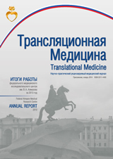
Translational Medicine

Advanced search

The «Translational medicine» journal is a peer-reviewed, open access journal aiming to bring together specialists working on the problem of health care, disease treatment, and increase of life expectancy. Implementation the concept of translational medicine requires close interaction of clinical and basic sciences, e.g. biology, physics, and chemistry to solve the specific clinical questions. Therefore, the objective of the journal is to assist Russian and foreign scientists carrying out their research in the field of translational medicine.

Established in 2015 Scientific and Educational Medical Cluster "Translational medicine" united Almazov National Medical Research Centre and the top rank Universities of Saint-Petersburg working in the field of drug and medical equipment development. The «Translational medicine» journal is the first journal covering the progress and achievements of the Cluster work.

The «Translational medicine» journal is included in the list of peer-reviewed scientific journals, not included in the international databases and citation systems, recommended to publish basic scientific results of dissertations for a degree of candidate of Sciences and doctor of Sciences on specialties: general biology, physiology, clinical medicine, preventive medicine, biomedical science.


Current issue

Vol 12, No 1 (2025)
View or download the full issue PDF (Russian)

PEDIATRICS

6-14 136
Abstract

Introduction. Hypertrophic cardiomyopathy (HCM) is the most common form of cardiomyopathy (CMP). Myocardial hypertrophy in children is associated with a large heterogeneous group of diseases with a high representation of non-sarcomeric causes. Purpose. The study compares diagnostic information from targeted next-generation sequencing (NGS) panels with different gene sets for HCM in children. Materials and methods. A molecular genetic study was conducted using two targeted panels (172 and 39 genes), followed by an assessment of their effectiveness. Results. The study included 53 children with HCM, the median age of which was 10 ± 3.5; 14.0 years. 13 children were examined using a large cardiac panel containing 172 genes. The remaining 40 patients were examined using a targeted panel for HCM containing 39 genes, while in 10 cases (25.0 %) genetic diagnostics was uninformative. Pathogenic and likely pathogenic variants in various genes were detected in 35 (66.0 %) patients. The informativeness of genetic diagnostics in the group of children under one year old was 60.7 %, in the group with onset at the age of over one year — 69.2 %. Conclusion. It is advisable to use an extended cardio panel for NGS for the purpose of genetic diagnosis of HCM in patients with the onset of the disease in the first year of life and small target panels for HCM in patients with the onset at an older age.

EDITORIAL

CELL, TISSUE, AND GENE THERAPY

17-26 96
Abstract

Background. In assisted reproductive technology (ART) programs, not all oocytes obtained during transvag inal puncture (TVP) are suitable for fertilization, as not all of them are at MII stage. Oocytes at germinal vesicle (GV) and MI stages are discarded because, after cumulus cells (CC) removal, their maturation and fertilization become impossible. Study proposes method for oocytes maturing by introducing extracellular vesicles (EVs) from follicular fluid (FF) into the perivitelline space. Objective. To evaluate the clinical feasibility of GV/MI oo cytes maturing technology without cumulus cells by injecting donor FF EVs under the zona pellucida. Materials and methods. FF (5 ml) was collected from 4 donors. EVs were isolated using sequential centrifugation. Portion of EVs was analyzed using Nanoparticle Tracking Analysis (NTA), while another portion was examined using transmission electron microscopy (TEM). Total of 53 immature oocytes were selected for the main group and 18 for the control group. Donor FF EVs were injected 4 hours after CC removal by introducing EVs suspension under zona pellucida. After 17 hours, oocyte maturity was assessed in both groups. Results. Oocytes maturation rate in the main group was statistically significantly higher than in the control group, indicating the potential effectiveness of the EV-IVM method for maturing GV-stage oocytes. Conclusion. Obtained data provide hope for the development of a new method for in vitro oocyte maturation.

27-36 81
Abstract

Background. Biological fluids contain diverse extracellular vesicle (EV) populations differing in size, origin, composition, and function. This study pioneers asymmetric depth filtration for isolating seminal plasma (SP) EVs, bypassing ultracentrifugation (current gold standard). Objective. Validate the novel asymmetric depth filtration method for SP EV isolation as an alternative to ultracentrifugation. Design and methods. Twelve samples of seminal plasma were collected from young men and patients of advanced reproductive age with mild teratozoospermia. EVs were isolated from the SP using the proposed method. Western blot analysis for vesicle membrane markers CD9, CD63, and CD81 confirmed the presence of SP EVs in the studied samples. Transmission electron microscopy further validated the presence of SP EVs. Morphological characteristics of SP EVs from men of different age groups were described. Quantitative characteristics of the SP EV samples were assessed using Nanoparticle Tracking Analysis (NTA). Results. The obtained particle size and concentration distributions revealed age-related changes in SP EVs: in the advanced reproductive age group, a shift in the dis tribution peak toward larger particles was observed compared to the peak distribution in the young men group. Conclusion. The results provide a basis for further investigation of SP EVs and exploration of their potential clinical applications in assisted reproductive technologies, such as in vitro fertilization programs.

37-50 79
Abstract

Relevance. Studies of the composition and properties of small extracellular vesicles (sEVs) are becoming more and more important due to their usefulness for the diagnosis and therapy of many pathologies. However, reliable universal methods of sEVs isolation and study have not been developed yet. Objective. To develop a modification of the method of sEVs isolation and to use the modified method for isolation of sEVs of neuronal origin. Materials and Methods. Using polyethylene glycol (PEG) precipitation at different pH and ionic strength values, sEVs were isolated from the blood of healthy volunteers. Immunoprecipitation was used to isolate sEVs of neuronal origin. Neuronal sEVs were biotinylated, and biotinylated proteins were identified by mass spectrometry. Results. As a result of this work, a modification of the PEG precipitation method at acidic pH values was developed, which allowed to increase the yield of neuronal sEVs several times. The surface proteins of sEVs were identified; these proteins were serum albumin, apolipoprotein B, complement system components C1r, C1q, C1s, C3 and immunoglobulin heavy chains. Conclusion. PEG precipitation under acidic conditions allows the isolation of more neuronal sEVs from serum than PEG precipitation did in neutral medium. At the same time, neuronal sEVs in blood most likely exist in complex with blood proteins, representing the so-called protein “corona”.

BIOENGINEERING AND BIOINFORMATICS

51-66 123
Abstract

Extracellular vesicles (EVs) are heterogeneous nanoparticles (30–500 nm) found in all biological fluids. The composition of EVs largely reflects the composition of the cells that secreted them. At present, EVs are widely used to search for diagnostic and therapeutic markers, and therefore studies of EVs composition and properties are becoming more and more in demand today. Despite the obvious potential of EVs in biomedical applications, the lack of unified methods for their isolation, analysis and quantitative measurement significantly limits scientific progress in this field. In scientific literature there are descriptions of dozens of methods for isolation and study of EVs, and often researchers do not present comparative advantages and disadvantages of the methods used. In this regard, a review of methods for the study of EVs becomes relevant. This review presents a comprehensive analysis of modern methods for the study of EVs. Each method has its own advantages and limitations affecting the yield, purity and characteristics of isolated vesicles, as well as the accuracy of the results obtained. The choice of the method for the isolation and analysis of EVs should be made considering the purpose and specificity of the study, since there is no universal approach at present. A thorough understanding of methodological nuances is critical for optimizing reproducibility and reliability in EV research.

67-79 120
Abstract

Background. Exosomes belong to the class of extracellular vesicles that are secreted by cells and circulate in biological fluids, mediating intercellular communication and signaling in various pathological processes, including modulation of cancer development and tumor microenvironment. Exosomes released by cancer cells at early stages of disease progression expressing specific membrane markers, which makes exosomes a promising biomarker of the disease by liquid biopsy. The main obstacle to the use of exosomes as markers of disease development is the lack of a convenient, inexpensive and rapid method for their isolation and detection. Objective. The aim of this study was to develop a platform for exosome isolation and detection of exosome protein membrane markers. Design and methods. In this work, magnetic nanoparticles functionalized with anti-EpCAM aptamer were developed for selective capture of tumor cell exosomes. The efficiency of magnetic nanoparticles was tested on exosomes derived from EpCAM-positive colon cancer cell line (HT29). To ensure high sensitivity of exosomes from pathologically cells, vesicles were pre-labeled with a lipophilic fluorescent dye (3,3’ dioctadecyloxacarbocyanine). Results. It was shown that magnetic nanoparticles bound with anti-EpCAM aptamer effectively captured exosomes from the HT29 culture medium. The detection limit of about 107 exosomes/ml was obtained using flow cytometry method. Conclusion. This study demonstrates an effective method for selective isolation of exosomes from cancer cell, which is promising for diagnostics and monitoring of oncological diseases.

80-93 99
Abstract

Exosomes are spherical extracellular nanovesicles of endosomal origin, whose function is to encapsulate part of the contents of the parent cells producing them and transport this content to the target recipient cells using biological fluids. Due to their properties, exosomes are considered as potential biological drug delivery systems. For medical purposes, exosomes are isolated from various natural sources. The use of each type of exosome for therapeutic purposes has its advantages and is associated to varying degrees with several biological (stability, immunogenicity, toxicity) and technical (production scaling-up, standardization of isolation protocols, drug loading) problems. Exosomes derived from human cells have significant potential as therapeutic drug (TD) delivery vehicles due to their endogenous origin. However, simultaneously with the delivery of TD, they can carry potentially dangerous biomolecules. Farm animal milk-derived exosomes and exosome-like plant-derived extracellular vesicles have enormous therapeutic potential in themselves and are safe as drug delivery vehicles. However, data on their effects on the human body are limited. Artificial exosomes created with the help of nanobiotechnology can overcome many of the technical limitations inherent in natural exosomes. The review discusses the strengths and limitations of different types of natural and artificial exosomes as drug delivery nanocarriers, as well as challenges associated with their implementation in clinical practice.

ALLERGY AND IMMUNOLOGY

94-107 98
Abstract

Antibiotic resistance is a rapidly escalating global healthcare crisis. Bacteria employ diverse strategies to evade both antibiotics and the host immune system. One of these tools is the use of bacterial extracellular vesi­ cles that mediate bacterial survival under antibiotic and immune stress. This review analyzes the impact of bacterial extracellular vesicles on antibiotic resistance and immune evasion. Bacterial extracellular vesicles of gram-positive and gram-negative bacteria have a number of structural differences, as well as different mechanisms for implementing their functions. Bacterial extracellular vesicles contribute to antibiotic resistance by acting as antibiotic targets, carrying resistance genes (horizontal gene transfer), and removing/degrading antibacterial agents. By implementing the strategy of evading immune surveillance, bacterial extracellular vesicles can participate in the formation of biofilms, provoke pro- and anti-inflammatory reactions, influence the secretion of cytokines by immune cells, creating more favorable conditions for colonization and development. Therefore, bacterial extracellular vesicles significantly contribute to bacterial survival under antibiotic and immune stress. However, the molecular mechanisms underlying bacterial extracellular vesi­ cles biogenesis and host responses to bacterial extracellular vesicles remain poorly understood, highlighting the need for further research to combat antibiotic resistance and bacterial infections.

Announcements

2025-05-12

Новый тематический выпуск журнала и специальный подкаст о внеклеточных везикулах

Журнал «Трансляционная медицина» представляет тематический выпуск, посвященный актуальным исследованиям внеклеточных везикул – ключевых инструментов межклеточных взаимодействий и перспективных терапевтических агентов современной медицины.

В номере представлены результаты фундаментальных и прикладных исследований отечественных специалистов в области разработки новых методов изучения внеклеточных везикул, а также клинические наблюдения в сфере гинекологии, андрологии и терапии. Особую ценность представляют обзорные и аналитические материалы, указывающие на перспективные направления дальнейших исследований и возможности практического применения полученных знаний.

Параллельно с публикацией тематического выпуска мы запустили специальный научно-популярный нейросетевой подкаст, который позволит глубже погрузиться в проблематику внеклеточных везикул и обсудить актуальные вопросы их исследования. Приглашаем всех заинтересованных специалистов ознакомиться с материалами выпуска.

Следите за новыми публикациями и будьте в курсе последних достижений современной медицины вместе с журналом «Трансляционная медицина»!

More Announcements...


Creative Commons License
This work is licensed under a Creative Commons Attribution 4.0 License.